👓 Beyond Flat Screens: Why 3D Matters in Microscopy
For over a century, microscopists have stared at flat 2D images of 3D worlds. Cells, tissues, and organisms exist in volumetric space — yet we've been forced to interpret them through flat projections. That's changing rapidly.
3D glasses and stereoscopic displays are revolutionising how researchers interact with microscopic data, offering:
- Depth perception — See cellular structures in true spatial relationships
- Intuitive navigation — Move through z-stacks naturally instead of clicking buttons
- Improved accuracy — Better judge distances, overlaps, and colocalisation
- Reduced eye strain — Natural stereoscopic viewing is more comfortable over long sessions
- Enhanced collaboration — Multiple viewers can share the same 3D experience
"The human brain is wired for stereoscopic vision. When we force researchers to interpret 3D structures from 2D projections, we're asking them to do cognitive gymnastics. 3D glasses remove that barrier."
🔬 Types of 3D Glasses and Display Technologies
1. Anaglyph Glasses (Red/Cyan)
The classic red-and-cyan glasses you might remember from 1950s cinema are still surprisingly relevant in microscopy:
- Extremely affordable (£2-5 per pair)
- Work with any standard display
- Can be used with monochrome SEM images when processed with anaglyph software
- Still used in many teaching labs for introductory stereoscopy
Note: While these glasses are primarily designed for entertainment (3D TV, movies, games), researchers have adapted them for viewing scientific anaglyph images including SEM micrographs. The colour accuracy is compromised — not suitable for fluorescence work where colour channels matter.
2. Active Shutter Glasses
These battery-powered glasses rapidly alternate between eyes in sync with the display:
- Full colour fidelity maintained
- Higher resolution per eye
- Used in professional stereoscopic microscopes
- Require compatible displays (120Hz+ refresh rate)
3. Polarised Glasses (Passive)
Lightweight and comfortable for extended wear:
- No batteries required
- Compatible with dual-projector or polarised display systems
- Used in surgical microscopes and training simulators
- Lower cost than active shutter systems
4. Autostereoscopic (Glasses-Free 3D)
The holy grail — 3D without glasses:
- Lenticular lens arrays direct different images to each eye
- Used in Light Field microscopes like the iMic3D
- Multiple viewers can see 3D simultaneously
- Technology improving rapidly but still expensive
5. Virtual Reality Headsets
The most immersive option:
- Full head tracking and spatial awareness
- Hand controllers for intuitive interaction
- Complete isolation from distractions
- Used in ConfocalVR and similar research platforms
📊 Technology Comparison
| Technology | Cost | Colour Accuracy | Comfort | Best For |
|---|---|---|---|---|
| Anaglyph | £2-5 | Poor | Good | Teaching, SEM |
| Active Shutter | £50-200 | Excellent | Moderate | Research, Fluorescence |
| Polarised | £10-30 | Good | Excellent | Surgery, Training |
| Autostereoscopic | £2,000+ | Good | Excellent | Collaboration, Displays |
| VR Headset | £300-1,000 | Good | Moderate | Immersive Analysis |
🧫 Applications in Cell Analysis
1. Colocalisation Studies
Determining whether two proteins or markers occupy the same spatial location is critical in cell biology. In 2D, this is ambiguous — overlapping projections might be separated in z. 3D glasses make spatial relationships immediately obvious.
Research from Stellenbosch University demonstrated that researchers using VR visualisation for colocalisation analysis achieved 40% faster region-of-interest selection with higher confidence ratings compared to traditional 2D interfaces.
2. Subcellular Structure Visualisation
Mitochondria, ER networks, and cytoskeletal elements are inherently 3D structures. Viewing them in stereoscopic depth reveals:
- True network connectivity
- Organelle-organelle interactions
- Spatial distribution patterns invisible in 2D
3. Cell Counting and Confluence
3D visualisation helps distinguish overlapping cells that appear merged in 2D projections, improving counting accuracy in dense cultures.
4. Tissue Architecture
In histology and developmental biology, understanding tissue organisation requires depth perception. Stereoscopic viewing of thick sections reveals:
- Layer relationships
- Invasion patterns
- Vascular networks
🤖 Wider Applications: Interacting with Machines
Beyond passive viewing, 3D glasses are enabling new ways to control and interact with microscopy systems:
1. Surgical Microscopy
Operating microscopes have long used stereoscopic vision — the surgeon's eyes are the 3D glasses. But new developments include:
- 3D heads-up displays — Overlaying diagnostic data in the surgeon's field of view
- Telemedicine — Remote surgeons viewing the same 3D image via glasses
- Training simulators — Medical students practising on 3D virtual tissue before touching real patients
2. Industrial Inspection
Manufacturing quality control uses stereoscopic microscopes for:
- Solder joint inspection (PCB manufacturing)
- Surface defect detection
- Dimensional measurements with depth accuracy
3. Telepresence Robotics
Researchers can control remote microscopes using 3D visualisation:
- Operate equipment in hazardous environments (biohazards, radiation)
- Share control with collaborators across the world
- Guide automated systems with human oversight
4. Augmented Reality (AR) Microscopy
Emerging technology overlays digital information onto the real microscope view:
- Measurement annotations floating in 3D space
- Real-time cell identification via AI
- Step-by-step protocol guidance for students
5. Human-Machine Interfaces (HMI)
3D glasses are becoming the interface between researchers and complex automation:
- Gesture control — Navigate z-stacks by waving hands
- Eye tracking — Focus on areas of interest automatically
- Voice commands — "Show me channel 2 at z-slice 45"
- Haptic feedback — Feel resistance when "touching" virtual cells
🌟 Case Study: ConfocalVR
Researchers at the University of California developed ConfocalVR, a system that converts confocal microscopy datasets into immersive VR environments. Users wearing VR headsets can:
- Walk around 3D cellular structures at true scale
- Measure distances by physically pointing
- Collaborate with colleagues in the same virtual space
- Export findings back to traditional 2D formats
The system reduced analysis time for complex 3D datasets by 60% while improving user satisfaction scores.
🔬 Leading 3D Microscopy Systems
The field of 3D microscopy is rapidly evolving with several innovative systems now available:
- Nanolive 3D Cell Explorer-fluo — Label-free 4D live cell imaging with integrated epifluorescence. Digital staining via refractive index eliminates need for dyes. Price: POA (typically £40,000-60,000)
- iMic3D Integral Microscope — Revolutionary light field microscope with glasses-free 3D display on Looking Glass monitors. Real-time spatial viewing for multiple viewers without glasses. Price: POA
- EVOS M5000 with 3D Visualisation — Advanced fluorescence system with 3D deconvolution and optional stereoscopic viewing accessories. Integrated cell counting with 3D depth rendering. Price: £12,000-15,000
👓 Getting Started: 3D Glasses for Your Lab
Budget Option: Anaglyph (£5-20)
Budget Option: Anaglyph (£5-20)
While primarily designed for entertainment (3D movies, TV, games), anaglyph glasses can be used in microscopy when images are processed into anaglyph format using software like ImageJ/Fiji:
- Works with any standard display
- Great for SEM images and monochrome datasets when converted to anaglyph format
- Students can experience depth perception affordably
- Compatible with ImageJ/Fiji anaglyph generation plugins
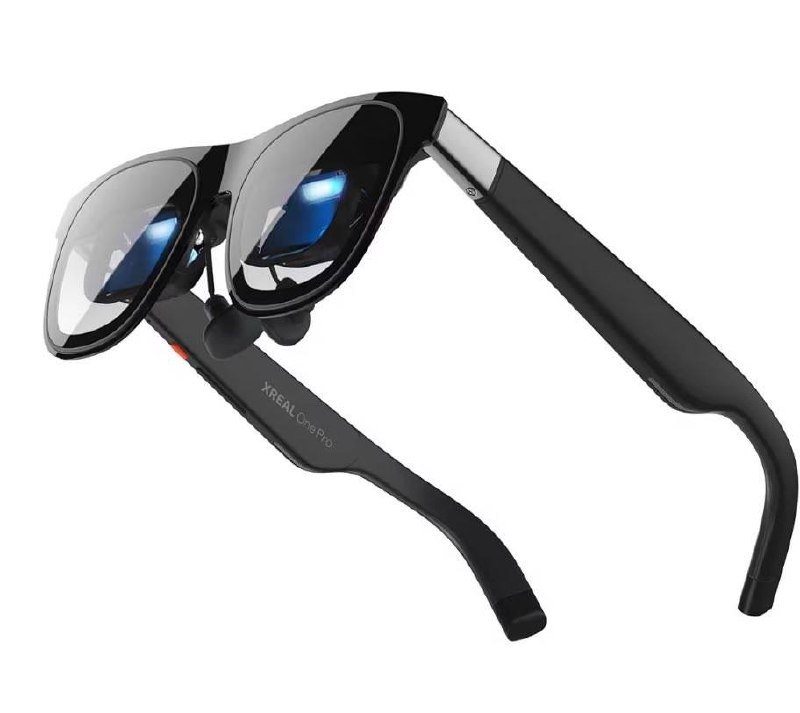
Note: These are different from AR glasses like XREAL — anaglyph glasses are simple colour-filtered lenses for viewing 3D content.
Mid-Range: Polarised or VR (£50-500)
For serious research applications:
- Passive polarised: Comfortable for long sessions, multiple users
- VR headsets: Full immersion with hand tracking
- Consider software compatibility for converting datasets to anaglyph format (ImageJ/Fiji plugins available)
Professional: Autostereoscopic Displays (£2,000+)
For collaboration and presentations:
- Looking Glass Factory displays
- Sony Spatial Reality Display
- Acer SpatialLab monitors
- Multiple viewers see 3D simultaneously
💡 Pro Tip
Before investing in expensive hardware, try software solutions that convert your existing microscopy datasets into anaglyph format. ImageJ/Fiji has plugins for anaglyph generation — a great way to test whether 3D viewing improves your workflow before spending money.
🔮 The Future: Where 3D Microscopy Is Heading
Near-Term (1-3 Years)
- Light field cameras becoming standard on research microscopes
- AI-assisted depth reconstruction from 2D images (computational 3D)
- Improved AR overlays for real-time guidance
- Cloud-based 3D sharing — send datasets to collaborators with guaranteed 3D rendering
Medium-Term (3-7 Years)
- Holographic displays — True 3D without glasses or headsets
- Brain-computer interfaces — Control microscopes with thought
- Digital twins — Virtual replicas of samples for infinite testing
- Remote telepresence — Operate any microscope on Earth from your office
Long-Term Vision (7+ Years)
- Direct neural interfaces — Experience microscopy data as synthetic vision
- AI co-analysis — Human and AI exploring datasets together in shared 3D space
- Haptic microscopy — "Feel" cellular textures and resistance
"The ultimate microscope won't be a device you look into — it will be a world you step inside. 3D glasses are the first step toward that future."
📊 Summary: Key Takeaways
- 3D glasses transform microscopy from passive viewing to immersive exploration
- Multiple technologies suit different budgets and applications — from £5 anaglyph to £50,000 light field systems
- Research shows 3D visualisation improves accuracy, speed, and user satisfaction
- Applications extend beyond viewing into machine control, telepresence, and AR guidance
- Accessible entry points exist — start with anaglyph or VR before investing in professional systems
🎯 Ready to See in 3D?
Whether you're a student exploring your first cells or a researcher analysing complex tissue architectures, 3D glasses offer a transformative upgrade to your microscopy workflow. Start simple with anaglyph glasses and discover a new dimension in your research.
Looking for AR glasses for gaming and media? Check out the XREAL One Pro featured at the top of this article.
📚 Further Reading
- Virtual Reality Assisted Microscopy Data Visualization (PMC)
- ConfocalVR: Immersive Visualization for Confocal Microscopy (PMC)
- VR Colocalisation Analysis in 3D Fluorescence Microscopy (PLOS One)
- High-Definition 3D Stereoscopic Microscope Display (Springer)
- Nanolive 3D Cell Explorer Official Site
- iMic3D Integral Microscope