Cell Culture Microscope Guide for Cell Painting Assays
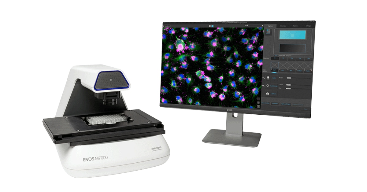
EVOS M7000 — Ideal for multi-channel Cell Painting assays. Image: Thermo Fisher Scientific
Compare the best inverted fluorescence microscopes for cell culture and tissue culture imaging, high-content screening, and automated morphological profiling.
🎨 What is Cell Painting?
Cell Painting is a high-content imaging assay developed at the Broad Institute that uses six fluorescent dyes to paint different cellular compartments. By capturing multi-channel images and extracting thousands of morphological features, researchers create a "fingerprint" of cellular states that can reveal how cells respond to genetic or chemical perturbations.
Hoechst 33342
Stains DNA in the nucleus. Reveals nuclear shape, size, and chromatin organization.
🛒 Buy Hoechst 33342Concanavalin A
Labels endoplasmic reticulum and Golgi. Shows protein synthesis and secretion pathways.
🛒 Buy Concanavalin AMitoTracker Deep Red
Labels active mitochondria. Reveals metabolic state and mitochondrial morphology.
🛒 Buy MitoTracker Deep RedWhy Cell Painting Matters
By combining six stains into a single multiplexed assay, Cell Painting creates rich morphological profiles that can distinguish between different cellular states, drug mechanisms, and disease models. This approach has been used to predict drug targets, identify novel therapeutics, and understand gene function at scale. The assay is compatible with most modern high-content screening platforms and open-source analysis tools like CellProfiler.
🔬 Best Cell Culture and Tissue Culture Microscopes for Cell Painting
Cell Painting requires a fluorescence-capable cell culture microscope or tissue culture microscope with at least 5 filter channels (DAPI, FITC, TRITC, Cy5, and a brightfield or phase contrast channel). Most researchers use inverted microscopes because they allow easy access to cell culture dishes, tissue culture flasks, and multiwell plates from above, while the objective sits below the stage.
🔬 Inverted Microscopes
Ideal for cell culture work. Objectives sit below the stage, leaving the top of dishes and plates unobstructed. Essential for live cell imaging and automated screening on multiwell plates.
🤖 Automated HCS Platforms
Systems like the Thermo EVOS M7000, Operetta CLS, or ImageXpress offer integrated environmental control, autofocus, and robotic plate handling for high-throughput Cell Painting at scale.
Key Specs for Cell Culture and Tissue Culture Microscopes
- Inverted design — for dish, flask, and plate access from above
- 5+ fluorescence channels — DAPI, FITC/GFP, TRITC, Cy5, plus brightfield
- 10x–40x objectives — 20x is the standard for Cell Painting
- Environmental chamber — CO₂, humidity, and temperature control for live cells and tissue cultures
- Motorized stage + autofocus — essential for automated multiwell and tissue culture screening
- High-resolution camera — ≥2MP monochrome sCMOS or CCD for quantitative imaging
🛒 Where to Buy Cell Painting Reagents
Purchase individual stains or complete Cell Painting kits from Thermo Fisher:
- Hoechst 33342 (Nuclear) — £89/10mL
- Concanavalin A (ER/Golgi) — £156/5mg
- SYTO 14 (RNA) — £178/5mL
- MitoTracker Deep Red (Mitochondria) — £234/20x50µg
- Phalloidin (Actin) — £156/300U
- WGA Alexa 555 (Membrane) — £189/1mg