🎬 Watch: Confluency Tool in Action
See how the real-time confluency algorithm works on the EVOS M3000 — from Thermo Fisher Scientific
▶
What is Cell Confluence?
Cell confluence is the percentage of the culture surface covered by cells. It's the most important metric in cell culture — it tells you when to passage, when to transfect, and when to run experiments.
Why Confluence Matters
- 70-80% confluence: Ideal time to passage — cells are healthy and actively dividing
- 90-100% confluence: Too late — cells stop dividing (contact inhibition), change phenotype
- 50-60% confluence: Too early — low yield, waste of media and reagents
- Transfection: 60-80% confluence gives best lipofection efficiency
- Drug treatments: Consistent confluence = reproducible IC50 values
The Problem with Manual Confluence Estimation
Most researchers estimate confluence by eye — looking down the microscope and guessing. This is surprisingly inaccurate:
| Issue | Impact | Result |
|---|---|---|
| Person-to-person variation | ±20% difference between researchers | Inconsistent passage timing, variable experiments |
| Small field of view | Judging entire flask from one microscope field | Misses edge effects, uneven growth |
| No documentation | No record of confluence at experiment start | Can't reproduce conditions or troubleshoot |
| Subjectivity | "Looks about 80%" vs precise measurement | Publication reviewers question methodology |
| Time consuming | 5-10 min per flask to check and estimate | 30 flasks = 2-3 hours/week just checking |
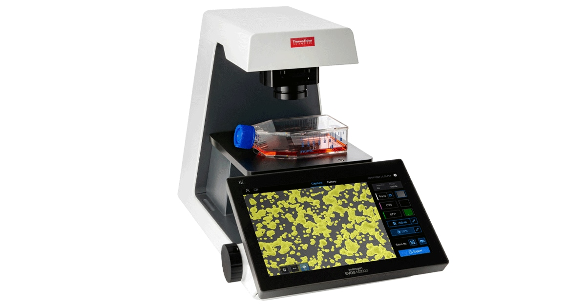
How the EVOS M3000 Confluency Tool Works
The EVOS M3000 confluency algorithm uses machine learning to automatically analyze the entire image and calculate precise confluence percentage in real-time.
Step-by-Step Process
- Capture image: Place flask on stage, select 4x or 10x objective
- Activate confluency tool: Tap the confluency button on touchscreen
- Real-time analysis: Algorithm instantly calculates % coverage
- Overlay display: Colored mask shows cell-covered vs empty areas
- Save result: Image + confluence % saved to USB or exported
🔬 Algorithm Details
- Phase contrast analysis: Detects cells without staining (live, non-invasive)
- Machine learning trained: On thousands of cell culture images across cell types
- Edge detection: Identifies cell boundaries vs background
- Real-time processing: Result in <2 seconds after capture
- Objective compensation: Calibrated for 4x, 10x, 20x magnifications
Benefits of Automated Confluence Detection
1. Reproducibility
- Same sample measured by different researchers = same result (±2% vs ±20% manual)
- Standardizes protocols across lab members and shift changes
- Year-over-year consistency — important for long-term studies
2. Time Savings
- Manual: 5-10 min per flask × 30 flasks = 3-5 hours/week
- Automated: 30 seconds per flask × 30 flasks = 15 minutes/week
- Time saved: 3-5 hours/week = 150-250 hours/year
- That's 4-6 weeks of researcher time recovered annually
3. Documentation
- Every measurement saved with timestamp and image
- Attach confluence data to experiment records
- Prove consistent cell state for publications
- Audit trail for GLP/GMP compliance
4. Better Science
- Transfect at exactly 70% every time = higher, reproducible efficiency
- Drug treatments at consistent confluence = valid IC50 comparisons
- No more overgrown cells with altered gene expression
- Detect growth rate changes early — flag problems before experiments fail
Real-World Applications
| Application | Confluence Target | Why It Matters |
|---|---|---|
| Routine passage (HEK293) | 80-90% | Maximum yield without contact inhibition |
| Transfection (lipofection) | 60-80% | Cells must be adherent but not overcrowded |
| Transfection (electroporation) | 80-90% | Higher confluence = better electroporation efficiency |
| Drug treatment (cytotoxicity) | 70-80% | Consistent starting density for valid comparisons |
| Wound healing assay | 100% | Monolayer must be completely confluent before scratch |
| Colony formation | 0% (single cells) | Cells plated at low density to form individual colonies |
| Differentiation (stem cells) | 60-70% | Lower confluence promotes differentiation vs proliferation |
💡 Pro Tips for Using the Confluency Tool
- Use 4x objective: Captures largest field for most representative measurement
- Measure 3 fields per flask: Center + two edges for average confluence
- Check calibration monthly: Run confluency on empty area = should read 0%
- Document edge effects: Confluence often lower at flask edges — factor into decisions
- Compare phase vs brightfield: Phase contrast gives sharper cell boundaries for analysis
- Export images: Save confluence overlay images for lab meetings and publications
🎬 Watch the Full Video
See the EVOS M3000 confluency tool in action:
▶ Watch on Thermo Fisher (2:52)Read EVOS M3000 Review →
🛒 Buy EVOS M3000 on Thermo Fisher UK →