🎬 Watch: Organoid Research with EVOS
See how EVOS imaging enables 3D cell culture analysis — from Thermo Fisher Scientific
The Challenge: Imaging 3D Cell Cultures
Organoids and spheroids present unique microscopy challenges:
- Depth: Structures 100-500μm thick — standard microscopes can't focus through entire organoid
- Heterogeneity: Different cell types at different depths require Z-stack imaging
- Time-lapse: Organoid growth takes days/weeks — need stable long-term imaging
- Quantification: Manual measurement of spheroid diameter is subjective and slow
- Multiple wells: Screening conditions across 96 wells with 3D structures
EVOS Solutions for Organoid Research
1. Z-Stack Imaging for 3D Reconstruction
- Capture images at multiple focal planes through the organoid
- Software reconstructs maximum intensity projection (MIP)
- View entire structure in single 2D image while preserving depth info
- Export Z-stacks for 3D rendering in external software (Imaris, Fiji)
Why 2D Deconvolution Matters for Organoids
After capturing Z-stacks through a 200-400μm organoid, you face a choice: how to present the data. Raw Z-stack slices show individual focal planes, but each plane contains out-of-focus blur from above and below. This is where 2D deconvolution becomes essential.
2D deconvolution applies a mathematical algorithm to each slice that removes out-of-focus light, restoring sharpness. For organoid research, this matters because:
- Sharper morphology: See cell boundaries and substructures that blur hides in raw images
- Better quantification: Automated cell counting and spheroid boundary detection work on deconvolved slices
- Reduced light dose: Deconvolution improves SNR without increasing excitation intensity — less phototoxicity for live organoids
- Publication quality: Deconvolved images meet journal standards without expensive confocal systems
- Speed advantage: 2D deconvolution processes each slice independently — faster than full 3D deconvolution, suitable for time-lapse organoid tracking
When to use 2D vs 3D deconvolution: For organoids < 300μm, 2D deconvolution on individual Z-slices followed by maximum intensity projection gives excellent results. For thicker organoids or when measuring 3D architecture, use 3D deconvolution that processes the entire Z-stack as a volume. The EVOS M7000 exports raw Z-stacks compatible with both approaches in Celleste or Fiji.
2. Automated Spheroid Analysis
- Auto-detect spheroid boundaries in brightfield or phase contrast
- Measure diameter, area, circularity automatically
- Track growth rate over time with time-lapse
- Compare drug effects across multiple wells simultaneously
- Export quantitative data with images for publications
3. Onstage Incubator for Long-Term Studies
- Maintain 37°C, 5% CO₂, humidity for days of imaging
- Capture time-lapse sequences every 30 min, 1 hr, or 6 hrs
- Monitor organoid maturation without removing from incubator
- Multi-well format — compare conditions in parallel
Applications in Organoid Research
| Application | EVOS Feature | Benefit |
|---|---|---|
| Tumor spheroid drug screening | 96-well scanning + growth analysis | Quantify IC50 in 3D models, not just 2D monolayers |
| Intestinal organoid budding | Time-lapse + Z-stack | Track crypt formation over 7+ days |
| Brain organoid development | Phase contrast + fluorescence | Monitor neural rosette formation with GFP markers |
| Liver organoid CYP induction | Multi-channel fluorescence | Measure CYP3A4-GFP reporter expression |
| Organoid-fibroblast co-culture | Cell counting by fluorescence | Quantify cell type ratios in mixed cultures |
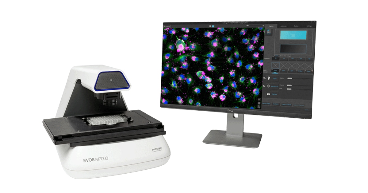
Recommended EVOS Configuration for Organoids
- EVOS M7000: Best for automated multi-well screening and analysis
- Objectives: 4x (overview), 10x (general), 20x (detail), 40x (substructure)
- Onstage Incubator: Essential for long-term time-lapse studies
- Celleste Software: Spheroid analysis module + Z-stack tools
- Plate formats: Ultra-low attachment 96-well plates (Corning, Thermo Fisher)
💡 Pro Tip: Best Settings for Organoid Imaging
- Use phase contrast for transparent spheroids (better than brightfield)
- Set Z-stack step size to 10-20μm for organoids 100-300μm diameter
- Use 2x2 binning for faster capture if resolution isn't critical
- For fluorescence: reduce excitation intensity to 20-30% to minimize phototoxicity during time-lapse
- Add anti-evaporation film for multi-day imaging to prevent edge effects
🎬 Watch the Full Video
See organoid imaging with EVOS in action:
▶ Watch on YouTubeRead EVOS M3000 Review →