🇬🇧 UK-focused independent guide. Plankton & Zoom reviews scanning microscopes used for virus plaque assays in UK virology, vaccine and gene-therapy labs. We do not sell systems. This guide explains what plaque assays are, compares colorimetric and fluorescent readouts, and matches workflows to the CellInsight CX5, EVOS M7000 and related platforms.
What Are Plaque Assays?
A plaque assay is the classic virology method for measuring infectious virus titre. A diluted virus sample is applied to a confluent layer of susceptible cells. After an incubation period, the virus kills or modifies small groups of cells, leaving visible clearings called plaques. Each plaque is assumed to originate from a single infectious particle, so the result is expressed as plaque-forming units per millilitre (PFU/mL).
Plaque assays sit at the heart of vaccine development, viral vector QC, antiviral screening, oncolytic virus production and biosafety testing. The format is simple — cells plus virus plus overlay — but the readout method has evolved from hand-counted crystal-violet dishes to automated fluorescent scanning on high-content microscopes.
For broader high-content and cell-imaging workflows, see our CellInsight CX7 review and EVOS M7000 review.
Colorimetric vs Fluorescent Plaque Readouts
| Readout type | Detection method | Microscope requirement | Best suited for |
|---|
| Crystal violet / staining | Absorbance-based colour change in infected/dead cells | Brightfield scanner or inverted microscope | Routine virus titration, teaching labs, fixed-endpoint assays |
| Immunostaining | Antibody-linked colour or fluorescence signal | Brightfield or fluorescence microscope | Virus-specific plaques, neutralisation assays |
| Fluorescent reporter virus | GFP, RFP or other fluorescent proteins expressed by engineered virus | Fluorescence scanning microscope | Live-virus tracking, high-throughput screens, early plaque detection |
| Live/dead viability dyes | Fluorescent dyes that stain intact or permeabilised cells | Fluorescence microscope or high-content imager | Cytopathic effect quantification, drug-toxicity panels |
Colorimetric assays are cheap and robust but rely on visible contrast, which can be subjective when plaques are faint or overlapping. Fluorescent readouts give higher contrast and can be counted earlier, before plaques become large enough to see by eye. They also multiplex easily with neutralisation markers or cell-health dyes.
Scanning Microscope Platforms for Plaque Assays
CellInsight CX5 High-Content Screening Platform
Five-channel laser-based plate scanner for automated plaque counting
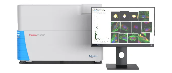
- Automated whole-well tiling and autofocus across 96/384-well plates
- Brightfield plus five fluorescence channels for multiplexed readouts
- Integrated HCS Studio software for object counting and size gating
- Ideal for virus neutralisation, antiviral screens and reporter-virus assays
The CX5 is built for plate-level statistics. It removes the manual stepping-stone stage by scanning every well on the same focal plane, normalising illumination and exporting plaque counts directly. For labs moving from 6-well teaching assays to 96-well screening, it is the logical upgrade.
Read CellInsight platform review
EVOS M7000 Imaging System
Automated inverted fluorescence microscope for development and low-throughput assays
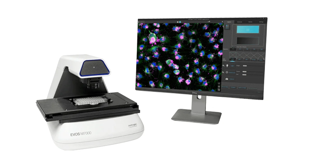
- Four-colour fluorescence plus transmitted light
- Automated stage, Z-stacking and time-lapse for plaque growth curves
- Cell counting and confluency analysis apps included
- Lower capital cost than a full high-content system
The EVOS M7000 is a practical bridge tool. It can image plaques in 6- to 24-well plates, capture publication images, and run reporter-virus time courses. It is the best fit when assay development precedes scale-up or when throughput stays below a few plates per week.
Read EVOS M7000 review
EVOS M5000 Imaging System
Five-channel benchtop fluorescence microscope for assay development
- Five fluorescence channels plus transmitted light
- No darkroom or eyepieces needed
- Automated cell counting and confluency apps
- Entry-level option for labs new to plaque imaging
Read EVOS M5000 review
How to Choose a Plaque Assay Microscope for a UK Lab
- Define the assay readout. Crystal violet needs brightfield only. Reporter viruses, immunofluorescence and live/dead dyes need fluorescence. If you mix readouts, buy a fluorescence-capable platform from the start.
- Pick the throughput format. 6- and 12-well dishes suit development. 96-well formats demand plate scanning. Multi-day time courses need environmental control or on-incubator imaging.
- Choose scanning hardware. High-content systems tile whole wells automatically. Benchtop inverted microscopes are cheaper and flexible but need more manual positioning.
- Set image-analysis rules. Normalise illumination, threshold by plaque size and intensity, and export PFU/mL. Lock the analysis protocol before switching routine assays from manual to automated counting.
- Validate against manual counts. Run a reference virus stock side-by-side. Establish equivalency limits and document them for GLP or GMP-adjacent work.
UK Microscope Buying Guide 2026
Frequently Asked Questions
What is a plaque assay used for?
Plaque assays measure infectious virus titre. A diluted virus sample is added to a cell monolayer; each infectious particle produces a visible clearing called a plaque. Results are reported as plaque-forming units per millilitre (PFU/mL).
What is the difference between colorimetric and fluorescent plaque assays?
Colorimetric assays use brightfield-visible stains such as crystal violet. Fluorescent assays use GFP reporter viruses, immunofluorescence or live/dead dyes and require a fluorescence scanning microscope.
Can the CellInsight CX5 run plaque assays?
Yes. The CX5 scans multiwell plates in brightfield and fluorescence, making it suitable for automated plaque counting, neutralisation assays and reporter-virus screens.
Can an EVOS microscope be used for plaque assays?
Yes. EVOS M5000 and M7000 systems image plaques in brightfield and fluorescence and are a good choice for assay development, lower throughput and labs that also need publication-quality cell images.
What magnification is best for plaque imaging?
2x to 4x objectives capture whole wells for counting, while 10x to 20x objectives resolve plaque morphology. High-content systems often use a low-magnification scan followed by a higher-magnification rescan of selected plaques.