🇬🇧 UK-focused independent guide. Plankton & Zoom compares fluorescence microscopes for neuroscience labs. We do not sell microscopes or broker quotes. This guide covers the systems most commonly used for calcium imaging, GFP reporter work, immunofluorescence, neurite outgrowth and high-content neural screening in the UK.
Why Fluorescence Microscopy Is Essential in Neuroscience
Neuroscience labs use fluorescence microscopy to see what brightfield and phase contrast cannot: calcium transients in living neurons, the localisation of synaptic proteins, the morphology of iPSC-derived neurons, and the response of neural cultures to compounds or genetic manipulations. The right fluorescence microscope lets researchers track dynamic processes, quantify structural changes and screen large numbers of conditions.
Choosing a system starts with the biological question. A lab doing fixed immunofluorescence of brain sections needs excellent sensitivity and stable illumination. A lab doing live calcium imaging needs speed, low phototoxicity and environmental control. A lab running drug screens on iPSC neurons needs automation, reproducibility and integrated analysis. These needs rarely all fit one microscope, so the goal is to match the platform to the dominant workflow.
For a broader overview of fluorescence microscope types and UK buying tiers, see our fluorescence microscope UK guide. For GFP and fluorescent protein imaging specifically, see Best Microscope for GFP Imaging UK.
Key Applications and Their Microscope Requirements
| Application | Channels needed | Critical features | Typical platform type |
|---|
| Live calcium imaging (GCaMP) | Green (FITC/GFP), sometimes red | Fast camera, low phototoxicity, environmental control, stable focus | Live-cell inverted fluorescence, spinning-disk confocal |
| GFP/RFP reporter tracking | Green, red, far-red | Sensitive detector, low bleaching, time-lapse capability | Inverted fluorescence, automated live-cell system |
| Fixed immunofluorescence (ICC/IHC) | Blue, green, red, far-red | Stable illumination, sensitive camera, easy multichannel merge | Inverted or upright fluorescence microscope |
| Neurite outgrowth assays | Blue, green, red | Automated stage, autofocus, image analysis software | Automated inverted fluorescence, high-content system |
| High-content neural screening | 5–7 channels | Plate handling, confocal option, integrated analysis, reproducibility | High-content screening platform |
| Brain slice / in vivo imaging | Green, red, sometimes multiphoton | Deep penetration, low scattering, upright or multiphoton optics | Two-photon / multiphoton microscope |
Most UK neuroscience labs doing cell culture work choose an inverted fluorescence microscope. Upright systems become relevant when the main workflow is acute brain slices or cleared tissue. Two-photon and light-sheet systems are specialised purchases usually handled through core facilities or dedicated imaging centres.
How to Choose a Fluorescence Microscope for Neuroscience Research
Follow this decision framework to avoid buying more microscope than you need, or under-specifying a system that will bottleneck your experiments.
- Define your biological question. Live calcium imaging, fixed immunofluorescence, GFP reporter tracking, organoid morphology and high-content screening each impose different constraints on speed, sensitivity and automation.
- Match the fluorescence channels. GCaMP and GFP need green excitation. RFP, mCherry and far-red antibodies need additional channels. Make sure the microscope has the LED or laser lines and the correct emission filters.
- Choose inverted or upright geometry. Inverted systems are easier for multi-well plates and live cell cultures. Upright systems with water-dipping objectives suit acute slices and cleared tissue.
- Check automation and throughput needs. Screens and large neurite-outgrowth assays need automated stage, autofocus and plate handling. Occasional imaging can use a simpler semi-automated system.
- Plan for UK support and software. Confirm warranty, field service, training, and whether image files export cleanly into Fiji/ImageJ, CellProfiler or your existing analysis pipeline.
UK Microscope Buying Guide 2026
Fluorophores and Filter Sets for Neural Imaging
Choosing fluorophores is as important as choosing the microscope. In neuroscience the most common combinations are:
- DAPI / Hoechst 33342: Nuclear counterstain for fixed ICC/IHC and cell-counting assays.
- GFP / FITC / GCaMP: Reporter proteins, calcium indicators and many primary antibodies.
- RFP / mCherry / Cy3: Second markers, co-culture labels and red-shifted reporters.
- Alexa Fluor 647 / Cy5 / far-red: Low-background antibodies and membrane dyes for three- and four-colour panels.
LED light sources give stable, long-lived illumination and are now standard on most modern systems. For very dim samples or fast dynamics, a sCMOS camera and solid-state excitation improve signal-to-noise and reduce photobleaching. For deeper imaging in tissue, two-photon excitation remains the gold standard.
Related Neuroscience Microscopy Workflows
Neuroscience labs often combine fluorescence microscopy with other imaging modes and assays. These Plankton & Zoom guides cover the surrounding workflow:
Frequently Asked Questions
What is the best fluorescence microscope for calcium imaging in the UK?
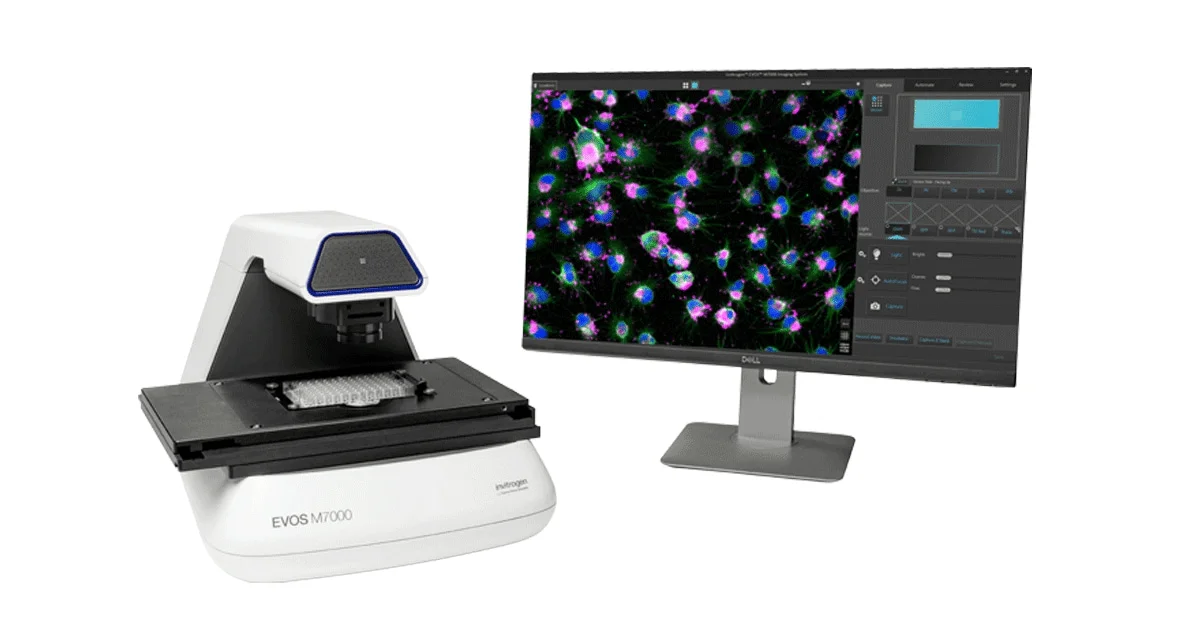
For cultured neurons and iPSC-derived models, the EVOS M7000 or a dedicated live-cell inverted fluorescence microscope with fast sCMOS camera and environmental control works well. For in vivo or slice calcium imaging, a two-photon or spinning-disk confocal system is usually needed.
Can the EVOS M5000 be used for neuroscience research?
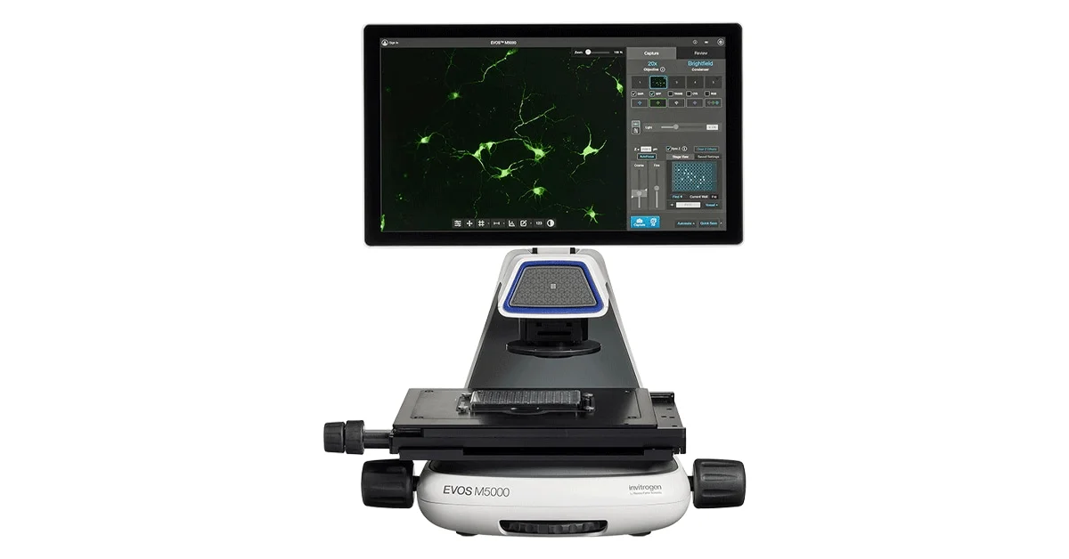
Yes. The EVOS M5000 is a five-channel inverted fluorescence microscope used in peer-reviewed neuroscience studies for neurite outgrowth, immunofluorescence, transfection and live-cell imaging of neural cell cultures.
Is CellInsight CX7 useful for high-content neuroscience screening?
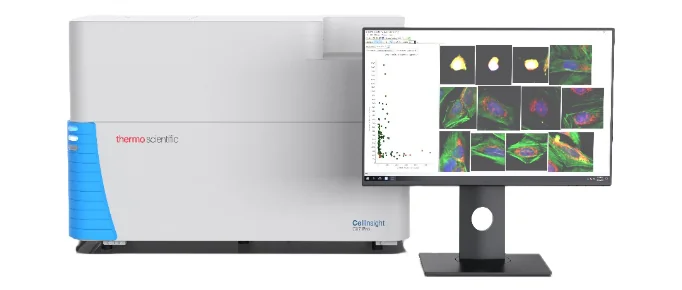
Yes. The CellInsight CX7 is a high-content screening platform with confocal and widefield options, suitable for morphological profiling of iPSC-derived neurons, neurite outgrowth assays and compound neurotoxicity screens.
What fluorophores are most common in neural imaging?
GCaMP and GFP for calcium and reporter imaging; RFP/mCherry for second markers; DAPI/Hoechst for nuclei; Alexa Fluor 647 or far-red dyes for antibodies and membrane markers.
Do I need confocal microscopy for neuroscience?
Not always. Widefield fluorescence is sufficient for monolayer cultures, neurite assays and most reporter work. Confocal or two-photon imaging becomes important for thick samples such as brain slices, organoids or in vivo preparations where out-of-focus light reduces image quality.
Compare UK fluorescence microscope tiers
Ready to shortlist a fluorescence microscope for your neuroscience lab? Plankton & Zoom compares EVOS, CellInsight, Zeiss, Leica and Nikon systems with UK tiers and authorised distributor links.
UK Microscope Buying Guide 2026
Ask a microscopy question