zenCELL owl Spheroid & Organoid Workflow UK Guide 2026
Label-free brightfield workflows for 3D cell cultures inside a standard CO₂ incubator.
Independent guide — Plankton & Zoom does not sell microscopes.
Why 3D Cultures Suit the zenCELL owl
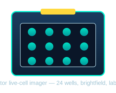
Spheroids and organoids spend most of their life inside a CO₂ incubator. Every time the door opens, temperature, humidity and gas equilibrium drift. The zenCELL owl removes that problem by living on the incubator shelf and imaging from below the plate. It is a label-free brightfield system, so there are no fluorescent reporters to photobleach, no fixing steps to interrupt kinetics, and no stains to wash away delicate extracellular matrix.
For UK labs running spheroid growth curves, organoid expansion or drug-response time-courses, this means continuous, multi-day data from a single plate position. We class the zenCELL owl in the Premium tier for dedicated incubator live-cell imaging.
3D Assays That Work Well on zenCELL owl
- Spheroid growth curves — measure projected area or confluency from seeding through compaction to plateau.
- Organoid expansion monitoring — track morphology changes during differentiation without disturbing Matrigel domes.
- Drug-response time-courses — capture compound-induced shrinkage, growth arrest or disaggregation over hours to days.
- Cytotoxicity assays — follow spheroid area loss as a label-free surrogate for viability when fluorescent endpoints are unavailable.
- Matrix invasion / migration — monitor brightfield area changes in embedded cultures, where the readout is area or boundary movement.
- Stem-cell aggregate QC — routine remote checks from the control PC to reduce incubator traffic.
Typical 3D Workflow on zenCELL owl
| Step | What the zenCELL owl does | Practical tip |
|---|---|---|
| 1. Plate cells | Waits inside incubator; imaging starts once scheduled. | Use a standard 24-well plate. Low-adhesion coatings or Matrigel domes should be centred over the well bottom. |
| 2. Set schedule | Captures each well at user-defined intervals from 1 minute to 24 hours. | For slow organoid growth, 6–12 hour intervals reduce file size while capturing morphology. |
| 3. Monitor growth | Records brightfield stacks for every well. | Avoid opening the incubator door during the first 24 hours to let thermal equilibrium stabilise. |
| 4. Add treatment | Continues imaging before and after compound addition for a continuous time-course. | Use rapid-pipetting workflows and note the exact frame time so treatment maps to image metadata. |
| 5. Analyse | AI confluency, projected area and growth-curve export. | Export CSV and import into Prism, Python or R for dose-response fitting. |
| 6. Export | PNG, JPG, AVI and CSV outputs plus ImageJ integration. | Save raw images in case reviewers ask for representative frames. |
3D Culture Platform Comparison for UK Labs
| Consideration | zenCELL owl | EVOS M7000 | CellInsight CX7 |
|---|---|---|---|
| Imaging mode | Label-free brightfield | Brightfield + fluorescence | Confocal + widefield fluorescence |
| Best for 3D | Growth curves and area kinetics | GFP reporters and z-stack morphology | High-content 3D screening |
| Incubator integration | Fits on incubator shelf | On-stage incubator chamber | External incubator options available |
| Plate capacity | 24 wells | Up to multiwell + slides | 96/384 high-content plates |
| Phototoxicity risk | Minimal — no excitation light | Low with optimised settings | Depends on laser/LED power and dose |
| Software complexity | Low — schedule, review, export | Medium — Celleste or vendor modules | High — HCS studio analysis |
| Qualitative tier | Premium dedicated incubator imager | Premium–core fluorescence system | Core-facility high-content platform |
UK Buying Guidance for 3D Culture Work
The zenCELL owl is supplied in the UK by InnoME GmbH and authorised scientific distributors. Because it is a dedicated incubator imager, final configuration, installation support and warranty terms affect the delivered package. Contact the manufacturer or a UK distributor for a current quote, demonstration and service coverage.
For labs that only need occasional brightfield checks, a standard inverted microscope may be sufficient. For labs needing fluorescent 3D reporters, confocal sectioning or high-content screening, consider the EVOS M7000 or CellInsight CX7 instead.
Frequently Asked Questions
Can zenCELL owl image through Matrigel or basement-matrix domes?
Yes, provided the dome is centred over the transparent well bottom. Brightfield imaging reads transmitted-light contrast through the plate base, so opaque inserts or very dense cultures may reduce visibility.
How do I turn brightfield area into a cytotoxicity metric?
Treat spheroid or organoid area as a surrogate for viable biomass. Plot normalised area over time after compound addition. A sustained drop relative to vehicle control suggests cytotoxicity or growth inhibition. Validate with an orthogonal viability assay before publication.
Can I use 96-well plates?
zenCELL owl is designed around a 24-well plate footprint. Check the latest datasheet for any adapter or format options if your workflow requires 96 wells.
Does the software measure spheroid diameter automatically?
The bundled software measures confluency and projected area. You can derive effective diameter from area assuming a circular projection, or export images to ImageJ for custom segmentation.