🇬🇧 UK-focused independent review. Plankton & Zoom does not sell microscopes or broker quotes. This guide explains what the EVOS S1000 does, where it fits in the Thermo Fisher imaging portfolio, and how it compares to the CellInsight CX5 high-content platform and conventional confocal systems for UK tissue-imaging workflows.
What Is the EVOS S1000 Spatial Imaging System?
The Invitrogen EVOS S1000 Spatial Imaging System is a slide-based fluorescence and brightfield microscope launched by Thermo Fisher Scientific in 2025 for spatial biology and tissue proteomics. Unlike the EVOS M5000 and M7000 — which are inverted cell-culture imagers — the S1000 is built to scan stained tissue sections on standard glass slides and generate multiplexed images with spectral unmixing.
Its headline claim is up to 9-plex fluorescence imaging in a single tissue section. That means researchers can label nine different proteins or markers simultaneously, capture the slide in one automated run, and computationally separate the fluorophores even when their emission spectra overlap. For cancer microenvironment studies, immuno-oncology and biomarker discovery, this kind of multiplexing turns a serial-section experiment into a single, spatially resolved dataset.
Why this matters to UK scientists: spatial proteomics is moving from specialist core facilities into mainstream cancer, immunology and pathology research. Systems that combine automated slide handling, spectral multiplexing and straightforward analysis software lower the barrier to entry and reduce the need to send every project to a spatial-omics core lab.
EVOS S1000 at a glance
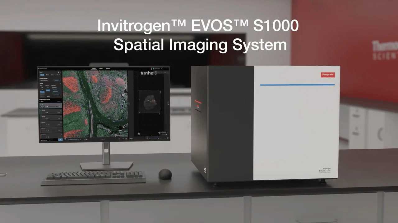
- Slide-based tissue imaging system
- Multiplex spectral fluorescence up to 9-plex
- Automated scanning and tiling
- Brightfield and fluorescence overlay
- Designed for spatial proteomics and tumour microenvironment research
View EVOS S1000 on Thermo Fisher UK
Technical Rationale: What the S1000 Does Well
1. Spectral multiplexing on fixed tissue
Traditional fluorescence microscopy is limited by filter sets: one excitation/emission pair per channel, and panels rarely exceed four or five markers before spectral overlap becomes unmanageable. Spectral systems capture the full emission profile of each fluorophore and use linear unmixing to recover individual signals. The S1000 applies this approach to tissue slides, enabling larger antibody panels without the cyclic staining protocols used by some CODEX or imaging mass-cytometry workflows.
2. Slide-based, not plate-based
The S1000 is optimised for standard histology slides. That makes it a natural addition for pathology, cancer research and translational labs that already produce FFPE or frozen sections. It does not require the multiwell plate format of high-content screening, so it can image clinical tissue arrays, tissue microarrays and serial sections with minimal workflow change.
3. Integration with the EVOS software ecosystem
Like other EVOS systems, the S1000 uses the familiar EVOS interface. Labs already running an EVOS M5000 or M7000 for cell culture do not have to learn a second acquisition environment to add tissue imaging. Images can be exported as standard TIFF for downstream analysis in QuPath, Fiji, CellProfiler or AI pipelines.
4. Throughput for small-to-medium panels
While it is not a whole-slide scanner, the S1000 is designed for targeted regions of interest rather than full-slide pathology screening. For research projects with defined marker panels — for example, CD3/CD8/PD-1/PD-L1 tumour immune profiling — it offers a middle ground between manual fluorescence microscopy and high-end whole-slide multiplex platforms.
EVOS S1000 vs CellInsight CX5 vs EVOS M7000
| Feature | EVOS S1000 | CellInsight CX5 | EVOS M7000 |
|---|
| Sample format | Glass slides / tissue sections | 96/384-well plates | Flasks, dishes, plates |
| Primary use | Spatial proteomics, multiplex IF | High-content screening | Live-cell imaging, assays |
| Fluorescence channels | Up to 9-plex spectral | LED fluorescence + brightfield | 5 fluorescence + transmitted light |
| Optical sectioning | Widefield / spectral | Widefield | Widefield with Z-stack |
| Live-cell imaging | No | Limited (plate-based) | Yes, incubator-compatible |
| Best for | Tissue sections, tumour microenvironment | Multiwell assays, screens | Cell cultures, time-lapse, Z-stacks |
The S1000 and CX5 are not competitors in the same workflow; they are complementary platforms. A UK cancer research group might use the S1000 to characterise marker expression across a tumour tissue bank, then use the CX5 or EVOS M7000 to run functional assays in cell lines or organoids derived from the same samples.
For a deeper comparison of EVOS systems, see our EVOS M7000 review and EVOS S1000 vs M7000 spatial biology guide.
Who Should Consider the EVOS S1000 in the UK?
- Cancer and immuno-oncology labs mapping immune infiltrates, checkpoint expression or stromal markers in tissue sections.
- Pathology and translational research groups adding multiplex immunofluorescence to existing H&E or IHC workflows.
- Core facilities that need a mid-tier spectral tissue imager between a manual fluorescence microscope and a high-end whole-slide scanner.
- Groups already invested in EVOS who want to extend the platform into tissue imaging without buying a separate software ecosystem.
Who should look elsewhere? Labs needing deep optical sectioning in thick cleared tissue, live-cell time-lapse, or fully automated high-throughput screening should consider confocal, light-sheet or CellInsight CX7/CX5 platforms instead.
Recent Publications and Resources
Multiplex tissue imaging and spatial proteomics are now mainstream in cancer biology. The references below explain why slide-based multiplex systems such as the EVOS S1000 are relevant and what technical standards the field is converging on.
Keren et al. (2018)
A structured tumour-immune microenvironment in triple negative breast cancer revealed by multiplexed ion beam imaging. Cell 174:1373-1387. doi:10.1016/j.cell.2018.08.039
View on DOI
Harms & Frankel (2023)
Multiplex immunohistochemistry and immunofluorescence: a practical update for pathologists. Modern Pathology 36:100197. doi:10.1016/j.modpat.2023.100197
View on DOI
Baccarani & Frangou (2024)
Spatial analysis by current multiplexed imaging technologies for the molecular characterisation of cancer tissues. British Journal of Cancer. doi:10.1038/s41416-024-02882-6
View on DOI
Mi et al. (2024)
Computational methods and biomarker discovery strategies for spatial proteomics: a review in immuno-oncology. Briefings in Bioinformatics. doi:10.1093/bib/bbae421
View on DOI
Lundberg & Borner (2022)
Spatial proteomics: a window into tissue architecture and function. Nature Reviews Molecular Cell Biology 23:375-380. doi:10.1038/s41580-021-00456-z
View on DOI
Gerdes et al. (2013)
Highly multiplexed single-cell analysis of formalin-fixed, paraffin-embedded cancer tissue. PNAS 110:11982-11987. doi:10.1073/pnas.1300136110
View on DOI
Frequently Asked Questions
What is the EVOS S1000 used for?
The EVOS S1000 is a slide-based spatial imaging system for multiplex fluorescence and brightfield imaging of tissue sections. It is used for spatial proteomics, tumour microenvironment mapping, immunofluorescence panels and marker colocalisation studies on fixed tissue slides.
How many fluorescence channels does the EVOS S1000 support?
Thermo Fisher advertises up to 9-plex spectral fluorescence imaging on the EVOS S1000, allowing researchers to unmix overlapping fluorophores and build large antibody panels on a single tissue section.
Is the EVOS S1000 a confocal microscope?
No. The EVOS S1000 is a widefield/spectral imaging system optimised for rapid multiplex tissue imaging. It produces high-resolution images but does not use a confocal pinhole for optical sectioning. For thin sections or cultured cells this is often sufficient; for deep tissue optical sectioning, a confocal or light-sheet system is more appropriate.
What is the difference between EVOS S1000 and CellInsight CX5?
The EVOS S1000 is a slide-based tissue imager for spatial proteomics and multiplex immunofluorescence. The CellInsight CX5 is a plate-based high-content screening system for automated multiwell assays. Choose the S1000 for tissue-section mapping and the CX5 for cell-based screening.
Can the EVOS S1000 do Z-stacks or live-cell imaging?
The EVOS S1000 is designed for fixed tissue slides, not live-cell or 3D culture work. For Z-stacks, time-lapse and live-cell growth curves, the EVOS M7000 is the better choice within the EVOS family.
What image analysis software works with EVOS S1000 images?
EVOS systems export standard TIFF images that can be opened in ImageJ/Fiji, QuPath, CellProfiler, Aivia, HALO and AI analysis pipelines such as CellPose. QuPath is particularly popular for multiplex immunofluorescence annotation and cell detection on tissue sections.
Compare the EVOS S1000 with UK alternatives
Need help deciding between EVOS S1000, CellInsight CX5 and other multiplex tissue-imaging platforms? Plankton & Zoom maps UK microscopy suppliers and compares systems for cancer, immuno-oncology and spatial proteomics workflows.
UK Microscope Buying Guide 2026
UK Microscopy Suppliers Map