EVOS S1000 Spatial Imaging System Review
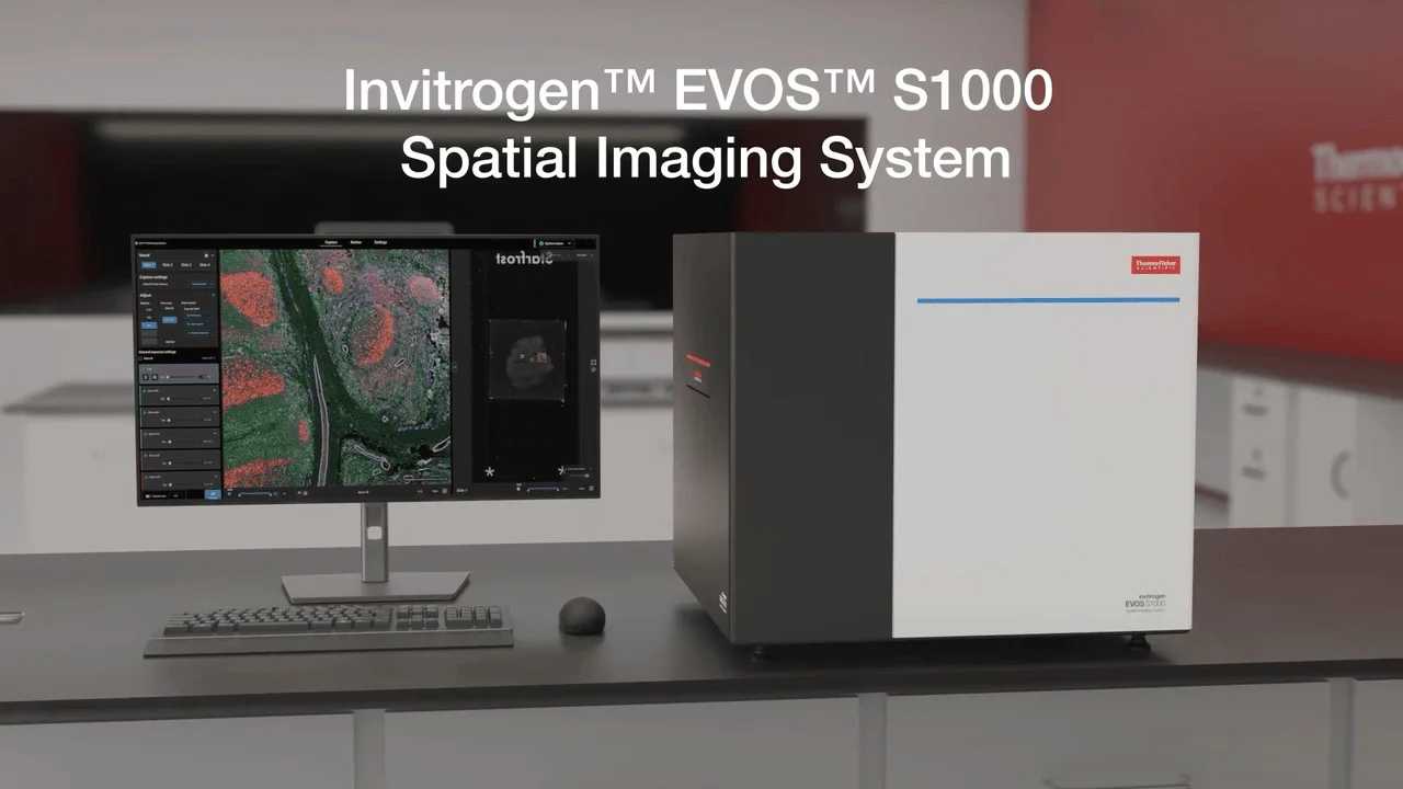
Image: Thermo Fisher Scientific
The EVOS S1000 Spatial Imaging System is Thermo Fisher's dedicated spatial biology platform for UK research and translational laboratories that need multiplex spectral fluorescence, tissue slide scanning and spatial proteomics on standard glass slides and coverslips.
🎬 Watch: Spectral Multiplex Fluorescence Imaging with the EVOS S1000
Thermo Fisher technology spotlight on practical panel design, spectral unmixing and spatial protein maps

Video: Thermo Fisher Scientific / Life Science Xchange
Key Features
- Multiplex Spectral Fluorescence: Up to 9-plex spectral fluorescence imaging with spectral unmixing for cleaner separation of overlapping fluorophores
- Tissue Slide Imaging: Optimised for imaging standard glass slides and coverslips with intact tissue architecture
- Automated Slide Scanning: Multi-region capture and automated XY scanning across tissue sections
- Spatial Proteomics: Map multiple protein markers in the same tissue section for immuno-oncology and translational research
- High-Resolution Scientific CMOS Camera: Captures detailed tissue morphology and single-cell information
- Integrated Acquisition and Analysis Software: Biology-focused interface from image capture to spatial annotation and export
Specifications
| Feature | Specification |
|---|---|
| Imaging Modes | Multiplex spectral fluorescence, Brightfield, Phase Contrast |
| Fluorescence Channels | Up to 9-plex spectral imaging (configuration dependent) |
| Camera | High-resolution scientific CMOS |
| Stage | Automated XY with multi-point navigation and slide scanning |
| Light Source | LED illumination with long lifespan and low heat |
| Sample Compatibility | Standard glass slides and coverslips |
| Software | Integrated EVOS capture, spectral unmixing and analysis tools |
| Data Export | TIFF, PNG, OME-TIFF, CSV metadata |
Who It's For
- Histology and pathology research laboratories
- Translational immuno-oncology groups
- Spatial biology and spatial proteomics researchers
- Cancer microenvironment and tumour immunology labs
- Core imaging facilities expanding into tissue workflows
EVOS Family Comparison
Not sure whether the S1000 is the right EVOS for your lab? Compare the full EVOS line-up:
- EVOS M3000 review - entry-level fluorescence imaging for cell culture
- EVOS M5000 review - professional four-colour fluorescence microscope
- EVOS M3000 vs M5000 vs M7000 UK 2026 - full three-way comparison
- EVOS M7000 review - automated live-cell and high-content imaging
- EVOS S1000 multiplex tissue imaging UK 2026 - dedicated guide to 9-plex spectral fluorescence and spatial proteomics
EVOS S1000 in Scientific Publications
The EVOS S1000 is referenced in the emerging spatial-biology literature. Applications include multiplex immunohistochemistry workflows, high-plex fluorescent labelling with spectral unmixing, tissue-section antibody validation, and spatial proteomics for translational research. A curated set of recent papers and resources is available in our dedicated research roundup.
EVOS S1000 Research Roundup → Spatial Biology Guide →
Workflows
Multiplex Immunofluorescence / Spatial Proteomics
Image multiple protein markers in a single tissue section using up to 9-plex spectral fluorescence. Spectral unmixing separates overlapping fluorophores so panels can be expanded beyond traditional filter-based systems, making it suitable for immuno-oncology phenotyping and tumour microenvironment studies.
Tissue Slide Scanning
Automated XY scanning captures multiple regions of interest across standard slides and coverslips. This walk-away capability supports high-throughput tissue imaging, whole-slide montages and repeat visits to the same ROI for longitudinal or multi-marker comparisons.
Spectral Unmixing
Spectral acquisition captures the full emission profile of each fluorophore, then software unmixes the signals. The result is cleaner channel separation and reduced bleed-through, which matters when building larger multiplex panels or working with autofluorescent tissue.
Pathology and Translational Research
By combining morphology, multi-marker protein localisation and spatial context on a benchtop instrument, the S1000 bridges traditional pathology imaging and higher-plex spatial platforms. It is positioned as an accessible entry point before committing to Xenium, MERFISH or mass-cytometry workflows.
UK Demo / Quote Guidance
The EVOS S1000 is available through Thermo Fisher Scientific UK and authorised UK distributors. For current product information, configuration options and to request a demonstration or official quote, contact the manufacturer directly.
- Thermo Fisher Scientific UK (Official Product Page) - Product details, specifications and quote/demo request
- Fisher Scientific UK - Distribution and support
Request Demo / Quote on Thermo Fisher →
Frequently Asked Questions
What is the EVOS S1000 used for?
The EVOS S1000 is a benchtop spatial imaging system for multiplex spectral fluorescence, tissue slide scanning, spectral unmixing and spatial proteomics on standard glass slides and coverslips.
How many fluorescent channels does the EVOS S1000 support?
It supports up to 9-plex spectral fluorescence imaging depending on configuration, enabling larger multiplex panels than traditional filter-based EVOS systems.
Can the EVOS S1000 do whole-slide imaging?
It performs automated multi-region scanning across tissue slides, allowing whole-slide montages and repeated visits to the same areas of interest.
Is the EVOS S1000 a spatial transcriptomics platform?
No. It is a spatial proteomics and multiplex immunofluorescence imaging system. For RNA-based spatial transcriptomics, labs typically use platforms such as 10x Genomics Xenium or Vizgen MERFISH.
Where can I request a UK demo or quote?
Contact Thermo Fisher Scientific UK or Fisher Scientific UK to arrange a demonstration or request an official quote.
What sample types work on the EVOS S1000?
It is designed for standard glass slides and coverslips with fixed tissue sections, cell smears and similar thin-sample formats used in histology and pathology workflows.