EVOS M3000 Review: Best Entry Level Fluorescence Microscope UK 2026
4.5/5 — Best Entry Level Fluorescence Microscope
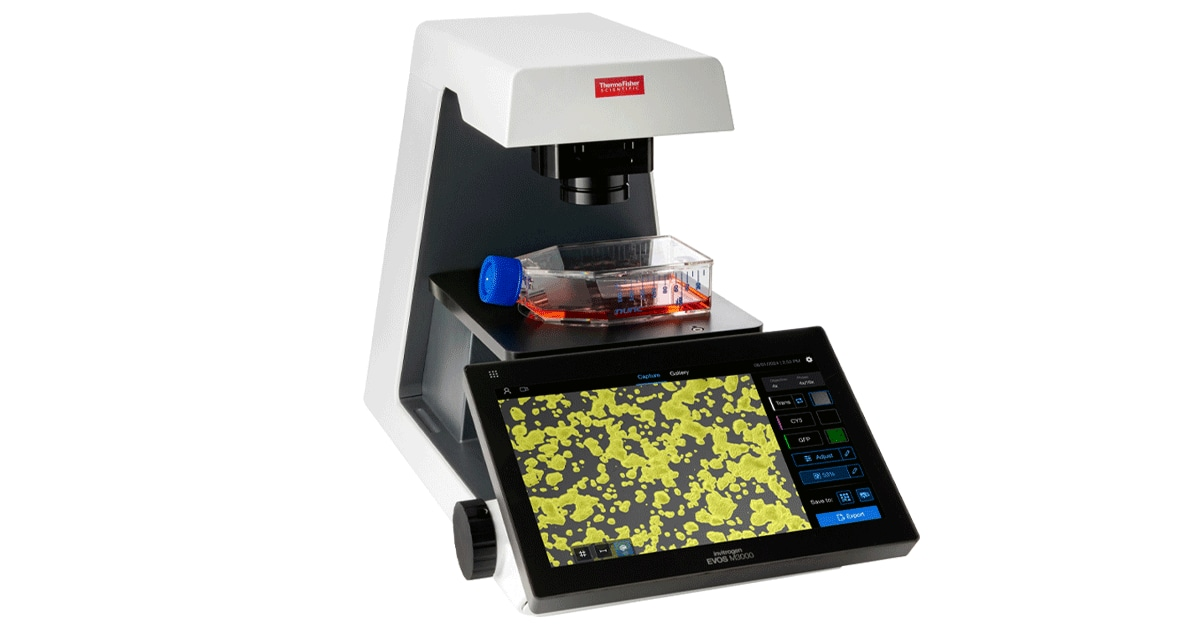
The EVOS M3000 is widely regarded as one of the best entry level fluorescence microscopes available in the UK market. Designed for teaching labs, research facilities, and routine cell culture work, this all-in-one system combines affordability with professional-grade imaging capabilities.
Key Features
- All-in-One Design: Integrated camera, monitor, and software — no external PC required
- LED Illumination: Long-lasting LED light source (50,000+ hours) with consistent output
- Touchscreen Interface: Intuitive controls reduce training time for students
- Cell Counting: Automated cell counting and confluence detection
- Live Cell Imaging: Compatible with onstage incubators for time-lapse studies
- USB Export: Easy image transfer for reports and publications
Specifications
| Feature | Specification |
|---|
| Imaging Modes | Fluorescence, Transmitted Light, Colour |
| Camera | Integrated Colour Camera |
| Monitor | Built-in LCD Touchscreen |
| Light Source | LED (50,000+ hour lifespan) |
| Software | On-board Image Capture & Analysis |
| Connectivity | USB, WiFi |
Best For
- Teaching laboratories
- Routine cell culture checks
- Multi-user facilities
- Labs transitioning to digital imaging
Where to Buy in the UK
The EVOS M3000 is available through:
View Product on Thermo Fisher →
🎬 EVOS M5000 Review with Video
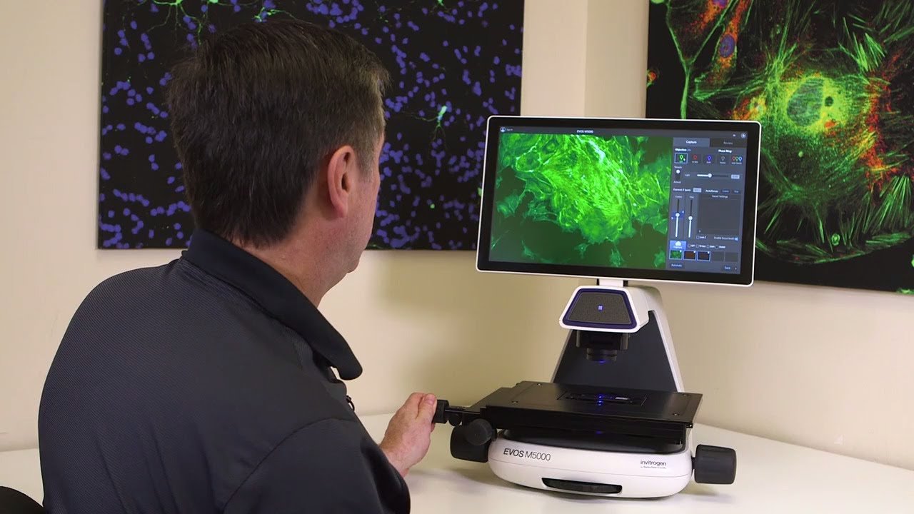
Looking for more advanced features? The EVOS M5000 adds automated multi-well scanning, time-lapse imaging, and advanced analysis software.
🎬 Watch Video & Read Review →
Vessel Compatibility Chart
The EVOS M3000 features Long Working Distance (LWD) objectives designed to image through plastic vessels. Use this chart to verify compatibility with your labware:
Multi-Well Plates
| Plate Type | Well Size | Recommended Objective | Imaging Quality |
|---|
| 6-well | 35 mm | 4x (overview) / 10x (detail) | ✅ Excellent — large field of view |
| 12-well | 22 mm | 10x / 20x | ✅ Excellent — optimal cell density |
| 24-well | 16 mm | 10x / 20x | ✅ Excellent — standard for assays |
| 48-well | 11 mm | 20x / 40x | ✅ Good — higher magnification needed |
| 96-well | 6.4 mm | 20x (recommended) | ✅ Good — single-cell resolution |
| 384-well | 3.2 mm | 20x / 40x | ⚠️ Challenging — small wells require precise positioning |
Flasks & Dishes
| Vessel | Dimensions | Recommended Objective | Notes |
|---|
| 35 mm Petri Dish | 35 mm × 10 mm | 4x / 10x / 20x | ✅ Standard vessel — full compatibility |
| 60 mm Petri Dish | 60 mm × 15 mm | 4x (overview) / 10x | ✅ Excellent for colony counting |
| 100 mm Petri Dish | 100 mm × 20 mm | 4x (overview) | ✅ Perfect for large-scale cultures |
| T-25 Flask | 25 cm² growth area | 4x / 10x | ✅ Standard small culture vessel |
| T-75 Flask | 75 cm² growth area | 4x / 10x | ✅ Most common routine culture vessel |
| T-175 Flask | 175 cm² growth area | 4x (overview) / 10x | ✅ Large-scale — excellent confluence overview |
Chamber Slides & Special Formats
| Vessel | Format | Recommended Objective | Best For |
|---|
| Chamber Slide (8-well) | Glass bottom option | 10x / 20x / 40x | ✅ Immunofluorescence, fixed cells |
| Microscope Slide | Standard 76 × 26 mm | 4x / 10x / 20x / 40x | ✅ Histology, tissue sections |
| Hemocytometer | Standard counting grid | 10x / 20x | ✅ Manual cell counting validation |
| Ibidi µ-Slide | Various formats | 10x / 20x / 40x | ✅ Live cell imaging, flow studies |
💡 Pro Tip: Objective Selection
4x Objective: Use for confluence measurements, colony overviews, and T-flask imaging. Large field of view captures entire wells.
10x Objective: The workhorse magnification. Perfect for routine cell culture checks, general morphology, and standard assays.
20x Objective: Ideal for 96-well plates, detailed morphology, and subcellular structures. Best single-cell resolution.
40x Objective: Use for detailed fluorescence imaging, organelle studies, and fine structural analysis. Smaller field of view requires stitching for large areas.
Long Working Distance (LWD) Objective Specifications
| Objective | Magnification | Working Distance | NA | Best Application |
|---|
| LWD Achromat | 4x | 20 mm | 0.13 | Confluence, colony overview |
| LWD Achromat | 10x | 7.8 mm | 0.25 | Routine cell culture |
| LWD Achromat | 20x | 6.5 mm | 0.40 | Detailed morphology |
| LWD Achromat | 40x | 3.5 mm | 0.60 | High-res fluorescence |
| LWD Phase | 10x | 7.8 mm | 0.25 | Phase contrast imaging |
| LWD Phase | 20x | 6.5 mm | 0.40 | Phase contrast detail |
Downloadable Validation Reports
Independent validation data demonstrating EVOS M3000 performance in real-world laboratory conditions:
🎯 Why Download These Reports?
For Lab Managers: Present to procurement committees as evidence for darkroom-free workflow transitions.
For Researchers: Compare SNR data against your current microscope to justify equipment upgrades.
For Teaching Labs: Use as training material to demonstrate modern fluorescence imaging capabilities.